
The human heart is an engineering marvel, beating roughly 100,000 times a day to pump vital oxygenated blood to every tissue in the body. However, the heart muscle itself relies on a dedicated network of pathways called coronary arteries to receive its own fuel supply. When this critical supply line is severely compromised or entirely blocked, the consequences are immediate, life-altering, and potentially fatal.
In everyday conversation, people frequently refer to this sudden crisis as a heart attack, but within healthcare settings, professionals use its formal designation: a myocardial infarction. Understanding what happens during this event requires looking past the sudden chest pain to examine cellular distress, complex electrical system anomalies, and rapid-response treatment pathways.
This comprehensive guide explores the multi-faceted nature of an acute myocardial infarction. We will dissect how it develops, how it shows up differently across populations, how doctors diagnose it using electrical tracings, and the immediate surgical and medical options used to save muscle tissue and lives.
What Is Myocardial Infarction in Medical Terms?
To establish a clear medical foundation, we have to look directly at the etymology of the phrase. When breaking down a myocardial infarction in medical terms, the phrase translates directly to the death of heart muscle due to a lack of blood supply.
The word “myocardial” combines myo (meaning muscle) and cardium (meaning heart). The word “infarction” refers to the tissue death (necrosis) resulting from a local lack of oxygen, typically caused by an obstructed circulation pathway. Therefore, the medical term myocardial infarction outlines a precise physiological event: a localized area of the heart muscle dies because its blood supply was cut off.
The Core Myocardial Infarction Definition
Looking at the strict clinical framework, the myocardial infarction definition centers on a prolonged imbalance between myocardial oxygen supply and demand. This imbalance leads to irreversible injury and death of myocardial cells.
The universal definition used by global cardiovascular organizations requires clinical evidence of myocardial necrosis in a setting consistent with acute myocardial ischemia. This evidence is usually gathered through specific blood tests, changes on a heart rhythm recording, or direct visualization of an artery blockage during emergency surgery.
Is Myocardial Infarction a Heart Attack?
A common point of confusion for many people reading health articles is the relationship between everyday terms and clinical terms. Put simply, a myocardial infarction is a heart attack. The two terms refer to the exact same underlying medical emergency.
While “heart attack” is the standard phrase used in common English, “myocardial infarction” (frequently abbreviated as MI) is the technical term utilized by physicians, nurses, and researchers. Writing or documenting a heart attack myocardial infarction ensures that both public-facing communication and clinical records align accurately.

Pathogenesis Myocardial Infarction and Plaque Development
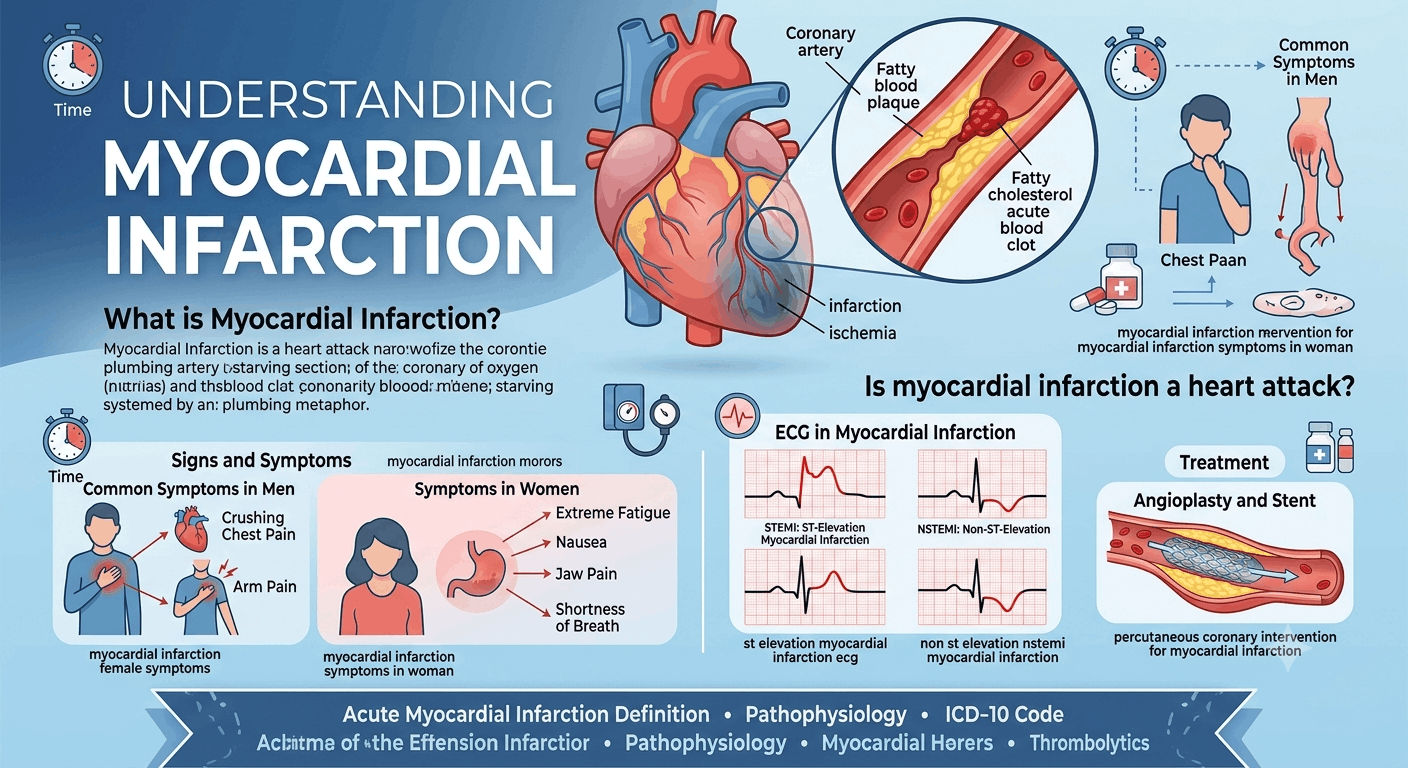
The structural decline leading up to an MI rarely happens overnight. The pathogenesis myocardial infarction process is deeply rooted in a chronic condition known as atherosclerosis, where fatty deposits, cholesterol, cellular waste products, and calcium build up along the inner linings of the coronary arteries.
Over decades, these materials organize into hard or soft structures known as atheromatous plaques. Soft plaques are particularly dangerous because they possess a thin fibrous cap covering a highly unstable, fatty core.
Detailed Myocardial Infarction Pathophysiology
The active myocardial infarction pathophysiology begins when one of these unstable plaques experiences a sudden physical disruption, such as a rupture, fissure, or erosion. When the plaque ruptures, the highly thrombogenic (clot-promoting) interior of the plaque is exposed directly to the circulating blood.
The body misinterprets this rupture as an active bleed and initiates a rapid clotting cascade. Platelets rush to the site, adhere to the broken plaque, and form a dense mesh of fibrin and red blood cells. If this resulting blood clot (thrombus) completely or partially seals off the inside of the artery, it causes an acute interruption of downstream blood flow.
Understanding Ischemia Myocardial Infarction Mechanics
Before cellular death actually occurs, the heart muscle enters a state of severe oxygen deprivation. This phase is known as ischemia. In the timeline of a myocardial infarction vs ischemia, ischemia represents the early, reversible stage of tissue distress.
During ischemia, cells are starved of oxygen but are still technically alive. If blood flow is restored quickly, the tissue can recover. However, if the ischemia continues uninterrupted for more than twenty to thirty minutes, the affected cells cross a point of no return, transitioning from temporary ischemia to permanent cell death, which completes the process of a true ischemia myocardial infarction.
Distinguishing Myocardial Infarction vs Cardiac Arrest
Another frequent point of confusion in medical media is the distinction between myocardial infarction vs cardiac arrest. While they are closely related and can happen during the same emergency, they are fundamentally different problems.
A myocardial infarction is a “plumbing” problem caused by a blocked pipe that cuts off blood flow, damaging a specific section of muscle. In contrast, a cardiac arrest is an “electrical” problem where the heart suddenly stops beating entirely, halting blood flow to the entire body. A plumbing problem (MI) can trigger an electrical failure (cardiac arrest), but they remain distinct clinical events.
Comparing Myocardial Infarction vs Heart Failure
It is also important to separate myocardial infarction vs heart failure. An MI is an acute, sudden event where muscle tissue dies due to a blocked artery. Heart failure, on the other hand, is a chronic, progressive condition where the heart muscle is too weak or too stiff to pump blood effectively to meet the body’s needs.
A severe or poorly treated myocardial infarction is one of the leading causes of chronic heart failure, as the scarred, dead muscle tissue left behind by an MI reduces the heart’s overall pumping capacity over time.
The Master Classification: Types of Myocardial Infarction
Not all heart attacks look or behave the same way under a microscope or on a hospital monitor. In clinical practice, healthcare providers categorize them into five specific types based on their underlying cause and the nature of the blood supply failure. The two most common variants encountered in emergency medicine depend on whether a coronary artery is completely blocked or only partially obstructed.
Defining Type 1 Myocardial Infarction
A classic Type 1 myocardial infarction represents the traditional heart attack model that most people are familiar with. It is caused by spontaneous plaque disruption followed by a blood clot that blocks blood flow. This type requires immediate medical or surgical intervention to clear the clot and restore flow to the dying muscle tissue before the damage becomes widespread.
Exploring Type 2 Myocardial Infarction
A type 2 myocardial infarction operates on a completely different mechanism. Instead of a sudden blood clot blocking an artery, a myocardial infarction type 2 occurs when a separate medical condition causes a severe mismatch between how much oxygen the heart muscle needs and how much it is actually receiving.
For instance, if a patient experiences extreme blood loss, a dangerously high heart rate, or a severe infection like sepsis, the heart muscle has to work much harder while receiving less oxygen-rich blood. This stress causes cell death even if the coronary arteries themselves are free of a sudden blood clot.
Silent Myocardial Infarction Pathologies
Alarmingly, a heart attack does not always present with dramatic, crushing chest pain. A silent myocardial infarction refers to an MI that occurs with very mild symptoms or no recognizable symptoms at all.
Patients experiencing a silent myocardial infarction might mistake their discomfort for mild indigestion, a pulled muscle, or a minor case of the flu. These events are often discovered weeks, months, or years later during a routine checkup or an electrical heart tracing. They are particularly common in elderly populations and individuals living with long-standing diabetes, as nerve damage can blunt the body’s ability to register cardiac pain.
Signs and Symptoms of Myocardial Infarction
Recognizing the signs and symptoms of myocardial infarction early is the single most critical factor in reducing tissue damage. When a classic event occurs, the body sends clear signals that the heart muscle is in severe distress.
The universal hallmark of an MI is chest discomfort, which patients often describe as a heavy pressure, squeezing fullness, or crushing pain in the center of the chest. This discomfort frequently radiates outward, travelling down the left arm, up into the jaw, or back between the shoulder blades.
Classic Myocardial Infarction Warning Signs
In addition to chest pain, there are several systemic myocardial infarction warning signs that indicate a medical crisis is underway. These include:
-
Sudden, unexplained shortness of breath (dyspnea)
-
Profuse, cold sweating (diaphoresis) that occurs without physical exertion
-
Overwhelming lightheadedness, dizziness, or a sudden drop in blood pressure
-
An intense, unexplained feeling of impending doom or severe anxiety
Myocardial Infarction Symptoms Men Experience
When analyzing how these events present across different populations, research shows that myocardial infarction symptoms men report tend to follow the classic textbook description. Men are highly likely to report sudden, intense, central chest pain that feels like a heavy weight sitting on their sternum. They also frequently experience radiating arm pain and profuse sweating, making the event easier for emergency medical teams to recognize immediately.
Breaking Down Myocardial Infarction Female Symptoms
For decades, public health models focused primarily on male presentations, leaving a gap in care for female patients. A myocardial infarction in woman populations often presents with subtler, less conventional signs.
While chest pain can still happen, myocardial infarction female symptoms are far more likely to focus on profound, unexplained fatigue that lasts for days before the event, severe shortness of breath, and a general feeling of weakness.
Understanding Myocardial Infarction Women Symptoms and Presentation
The variations in myocardial infarction women symptoms make these events harder to spot without clinical training. Women frequently experience discomfort that is localized entirely outside the chest, such as an aching pain in the lower jaw, neck, or upper back.
Because these symptoms lack the classic, crushing central chest pressure, women often delay seeking medical care, which increases the risk of complications.
Identifying Symptoms of Myocardial Infarction in Females
When evaluating the specific symptoms of myocardial infarction in females, gastrointestinal distress is another major factor. A female myocardial infarction frequently presents with severe nausea, vomiting, or a burning pain in the upper stomach that mimics acid reflux or indigestion.
Any woman experiencing unexpected, unexplained upper body discomfort combined with nausea and shortness of breath should be evaluated for a cardiac event immediately rather than waiting for classic chest pain to appear.
Myocardial Infarction in Young Populations
While heart attacks are widely viewed as conditions affecting older adults, a young myocardial infarction is becoming increasingly common in modern healthcare. An MI occurring in adults under the age of fifty is often tied to unique risk factors, including strong genetic predispositions to high cholesterol, a history of heavy smoking, high stress levels, or the use of illicit stimulants.
Additionally, rare cases involving the sudden tearing of a coronary artery wall (spontaneous coronary artery dissection, or SCAD) can cause an acute MI in young, otherwise healthy individuals.
High-Profile Cases: Athlete Founder of Investment Bank Died of Myocardial Infarction
The reality that an MI can strike individuals who appear to be in peak physical condition was brought into the public eye by a tragic, widely reported news story. Media reports noted that an athlete founder of investment bank died of myocardial infarction suddenly, causing shockwaves across both the financial and athletic communities.
This tragic case highlighted how underlying, undiagnosed cardiovascular disease or plaque vulnerabilities can exist silently even in individuals who maintain highly active lifestyles.
The Intersection of Sports and Finance: Former Footballer Founder of Investment Bank Died Myocardial Infarction
Further investigations into this high-profile case revealed that the individual was a former footballer founder of investment bank died myocardial infarction. This background as an elite, competitive athlete underscored an essential medical truth: past athletic performance and outward physical fitness do not provide complete immunity against coronary plaque rupture. It emphasized the critical need for comprehensive cardiovascular screenings, regardless of a person’s athletic history or current fitness level.
Diagnostic Medicine: The Myocardial Infarction ECG
When a patient arrives at an emergency department with a suspected heart attack, the most important diagnostic tool is the electrocardiogram, widely known as a myocardial infarction ecg or ekg.
An electrocardiogram myocardial infarction tracing records the electrical signals traveling through the heart muscle. When a region of the heart is starved of oxygen, its cells cannot maintain their normal electrical charges. This electrical disruption creates distinct, measurable patterns on the paper tracing, allowing doctors to confirm an MI within seconds.
Reading a Myocardial Infarction on an ECG
When a physician reviews a myocardial infarction on an ecg, they look closely at the different segments of the heart’s electrical wave profile. The electrical cycle consists of a P wave (atrial activation), a QRS complex (ventricular activation), and a T wave (ventricular recovery).
The flat line connecting the end of the QRS complex to the beginning of the T wave is known as the ST segment. Deviations in this specific segment are what doctors use to determine the severity and location of an active heart attack.
Understanding St Elevation Myocardial Infarction
The most severe category of heart attack is a st elevation myocardial infarction, commonly abbreviated as a STEMI. A STEMI occurs when a coronary artery is completely blocked by a blood clot, cutting off all blood flow to an entire region of the heart muscle.
On an ekg, this total blockage causes the ST segment to lift significantly above its normal flat baseline. A st-elevation myocardial infarction ecg profile requires immediate emergency treatment to open the blocked vessel before the entire thickness of the heart wall dies.
Analyzing Non St Elevation Myocardial Infarction
If the coronary artery is only partially blocked, or if the collateral blood vessels manage to provide a small amount of backup flow, the patient may experience a non st elevation myocardial infarction, known as an NSTEMI.
Because the blockage is incomplete, a non st wave elevation myocardial infarction does not cause the ST segment to lift on an ECG. Instead, it may cause the ST segment to depress downward or cause the T wave to flip upside down. Despite the lack of ST elevation, an NSTEMI still causes irreversible muscle damage and requires urgent medical management.
Anatomical Locations: Anterior Myocardial Infarction
The specific coronary artery that is blocked determines which part of the heart muscle suffers damage. An anterior myocardial infarction occurs when there is an obstruction in the Left Anterior Descending (LAD) coronary artery, often called the “widowmaker” because it supplies blood to the front wall of the heart.
An anterior myocardial infarction on ecg is visible across the chest leads (V1 through V4) and carries a high risk of complications because the front wall of the left ventricle is responsible for the majority of the heart’s pumping power.
Identifying Inferior Myocardial Infarction
An inferior myocardial infarction occurs when the blockage is located in the Right Coronary Artery (RCA), which supplies blood to the bottom (inferior) wall of the heart.
When diagnosing an inferior wall myocardial infarction, doctors look for specific changes in the electrical leads that face the bottom of the heart (leads II, III, and aVF). An ecg inferior myocardial infarction pattern requires careful management because damage to the inferior wall can cause a severe drop in blood pressure and disrupt the heart’s natural pacemaker cells.
Other Anatomical Variations: Posterior and Lateral Infarctions
Heart attacks can also impact other regions of the ventricular walls. A lateral myocardial infarction involves the left side wall of the heart, usually caused by a blockage in the Left Circumflex artery. A septal myocardial infarction damages the muscular wall that separates the left and right sides of the heart.
More complex cases include a posterior myocardial infarction, which affects the back wall of the heart, or an inferoposterior myocardial infarction ecg pattern, where an obstruction damages both the bottom and back walls simultaneously. Because standard chest leads do not sit on a patient’s back, a posterior myocardial infarction on ecg often requires placing extra electrical leads on the patient’s back to catch the injury.
Laboratory Diagnostics: Myocardial Infarction Creatine Kinase and Troponin
While an ECG provides an immediate visual look at the heart’s electrical activity, blood tests are essential to confirm that muscle tissue has actually died. When myocardial cells crack open and die during an infarction, they release internal proteins into the bloodstream.
Historically, measuring myocardial infarction creatine kinase (specifically the CK-MB fraction) was the gold standard for tracking this damage. In modern medicine, while creatine kinase is still monitored in specific contexts, it has been largely replaced by highly sensitive cardiac Troponin tests, which can detect incredibly small amounts of heart muscle injury with extreme precision.
Understanding Clinical Timeframes: Myocardial Infarction Time
In the management of a cardiovascular crisis, myocardial infarction time is the single most critical factor determining a patient’s long-term recovery. Cardiologists frequently use the phrase “time is muscle” to emphasize this urgency.
If a blocked artery can be opened within the first ninety minutes of symptom onset—a window known as the “golden hour”—a significant portion of the threatened heart muscle can be saved. As the clock ticks past this window, the area of permanent scarring grows larger, permanently reducing the heart’s long-term pumping capacity.
Emergency Interventions: Percutaneous Coronary Intervention for Myocardial Infarction
For a definitive, life-saving st elevation myocardial infarction treatment, the gold standard intervention is percutaneous coronary intervention for myocardial infarction, commonly referred to as a PCI or an emergency angioplasty.
During a PCI, an interventional cardiologist inserts a thin, flexible tube called a catheter into a blood vessel in the wrist or groin and guides it up to the blocked artery in the heart.
Acute Myocardial Infarction Drugs and Thrombolytics
If a patient arrives at a hospital that lacks a specialized cardiac catheterization lab for a PCI, or if transfer to such a facility will take too long, the medical team must rely on emergency medications. In these time-critical scenarios, doctors use thrombolytics for myocardial infarction.
Thrombolytics are powerful, clot-dissolving medications administered through an IV. These medications work by breaking down the fibrin mesh of the blood clot, acting as a chemical drain opener to quickly restore blood flow to the starving muscle tissue.
Long-Term Myocardial Infarction Medication and Management
Once the immediate blockage is cleared, the treatment focus transitions to preventing another event and helping the damaged heart heal. Long-term myocardial infarction medication therapies generally include a combination of several life-saving drug classes:
-
Antiplatelet medications: Medications like aspirin acute myocardial infarction protocols and P2Y12 inhibitors keep platelets slippery to prevent new clots from forming on stents.
-
Beta-blockers: These reduce the heart rate and blood pressure, lowering the heart’s workload and protecting it from dangerous rhythm disruptions.
-
ACE inhibitors: These help prevent the heart muscle from stretching out of shape as it heals around the new scar tissue.
-
Statins: High-dose statins stabilize existing plaques throughout the body and aggressively lower cholesterol levels.
Medical Coding and Documentation: Myocardial Infarction ICD 10
Behind the clinical care, healthcare systems rely on standardized codes to track diseases, process insurance, and maintain accurate records. The global standard for this documentation is the International Classification of Diseases, Tenth Revision, or myocardial infarction icd 10.
Using specific codes ensures that the exact nature, severity, and location of the heart attack are documented uniformly across different medical systems worldwide.
Common Codes: ICD 10 Code for Myocardial Infarction
When an administrative team inputs data, they must select the precise icd 10 code for myocardial infarction that matches the physician’s diagnosis. The primary coding category begins with I21.
An acute STEMI of the anterior wall is coded as I21.0, while an acute STEMI of the inferior wall is coded as I21.1. A standard NSTEMI is coded under I21.4. If a patient has a past history of a heart attack but is not currently having one, coder teams use a separate code, history of myocardial infarction icd 10 (which maps to I25.2), to show that healed scar tissue is present.
Final Word on Myocardial Infarction Prevention and Recovery
A myocardial infarction remains one of the leading causes of mortality and long-term disability worldwide, yet it is a condition where swift action, education, and lifestyle modifications can alter the course of the disease. Recognizing that an MI is a true plumbing emergency—distinct from electrical cardiac arrests or chronic heart failure—empowers individuals to act fast when warning signs appear.
By understanding that symptoms can look completely different in women compared to men, managing classic risk factors like high blood pressure and cholesterol, and supporting rapid emergency response systems, we can protect heart muscle, improve long-term recovery outcomes, and save countless lives.
Frequently Asked Questions
What is the main difference between an NSTEMI and a STEMI heart attack?
A STEMI (ST-Elevation Myocardial Infarction) happens when a coronary artery is completely blocked by a blood clot, causing widespread, immediate damage through the entire thickness of the heart wall, which shows up as an elevated ST segment on an ECG. An NSTEMI involves a partial or temporary blockage, meaning the muscle damage is less immediate and does not cause ST elevation on an electrical tracing, though it still causes cell death confirmed by blood tests.
Why do women experience different heart attack symptoms than men?
Women often experience atypical heart attack symptoms due to differences in how cardiovascular disease develops in female bodies. Women are more likely to have blockages in smaller branching blood vessels (microvascular disease) rather than a single major plaque rupture in a large artery. This distinct pathology often causes diffuse symptoms like extreme fatigue, shortness of breath, jaw pain, and nausea instead of classic, crushing central chest pain.
What should I do immediately if I suspect someone is having a myocardial infarction?
If you suspect someone is having a myocardial infarction, call emergency services immediately. Do not attempt to drive the person to the hospital yourself unless there is no other option, as emergency medical personnel can begin life-saving diagnostics and treatments the moment they arrive. While waiting for help, have the person sit down, stay calm, and chew a full-strength adult aspirin if they are not allergic.
Can a person fully recover after experiencing a myocardial infarction?
Yes, many individuals go on to live long, active lives after a myocardial infarction, provided they receive prompt treatment to minimize muscle damage. Long-term recovery relies on strict adherence to prescribed medications, participating in a structured cardiac rehabilitation exercise program, making heart-healthy dietary changes, and managing underlying risk factors like smoking, high blood pressure, and stress.